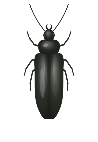
External Anatomy of Insects: Head, Thorax, Abdomen and Appendages
How an insect’s body is built, from the plates of its outer skeleton to the jaws, wings and breathing holes that make each order recognizable.
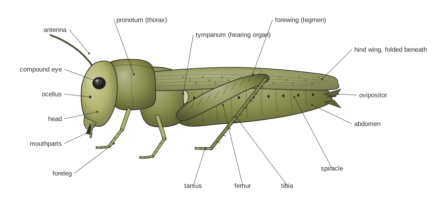
An insect’s body has three regions. The head carries the antennae, eyes and mouthparts. The thorax carries three pairs of jointed legs and, in most adults, one or two pairs of wings. The abdomen holds most of the digestive and reproductive organs and many of the breathing openings. All of it is wrapped in a jointed outer skeleton, the exoskeleton.
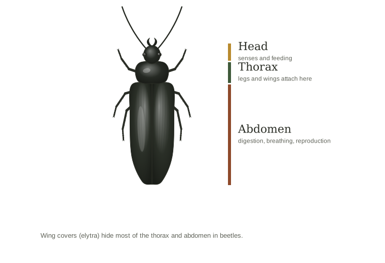
Knowing these parts is the quickest way to place an insect in its order. Wing type, antenna shape, mouthparts and the shape of the waist separate beetles from bugs, ants from termites and bees from flies. This guide moves from the outside covering inward to each body region, then closes with a practical checklist for identification.
The exoskeleton and body segments
Insects have no internal bones. Support comes from the cuticle, a layered outer skeleton made by a single sheet of skin cells (the epidermis) just beneath it.
The cuticle has two main parts:
- Epicuticle: a thin outer layer with a film of wax that keeps the insect from drying out. In many insects a cement layer covers the wax and protects it from wear.
- Procuticle: a thicker layer of chitin fibers set in protein. Where the outer part of it hardens, it forms a stiff exocuticle over a softer endocuticle.
Sclerites, sutures and membranes
Shortly after each molt, proteins in parts of the cuticle bond together and harden. The hardened plates are called sclerites. Between them, the cuticle stays soft and flexible. These membranes are rich in resilin, a rubbery protein, and they let the body bend at joints and between segments.
The cuticle also folds inward in places to form internal ridges and braces for muscles. From outside, a ridge shows as a groove called a suture, and a finger-like brace shows as a small pit.
Tergites, sternites and segmentation
Insects are built from a row of segments, a pattern easiest to see on the abdomen. Each segment has a hard upper plate (tergum or tergite) and a hard lower plate (sternum or sternite). On the abdomen, these are joined along each side by a soft pleural membrane. Soft intersegmental membranes connect one segment to the next, and the front edge of each segment tucks under the one ahead. This telescoping lets the abdomen stretch after a meal or as eggs develop.
Molting
A hardened exoskeleton cannot grow, so a young insect must replace it to get bigger. Hormones start the process. The skin cells pull away from the old cuticle (apolysis) and build a new one underneath it. The insect then splits the old skin and climbs out (ecdysis). The new cuticle is soft and pale at first. The insect swells it with air or fluid before it hardens and darkens.
Each stage between molts is called an instar. Most insects stop molting once they become adults. Silverfish and their relatives are the exception and keep molting throughout life. The shed skins left behind are often the first clue to an infestation of bed bugs, cockroaches or carpet beetles. For how molting fits into the life cycle, see simple and incomplete metamorphosis.
The head
The head is a hard capsule that holds the brain, the mouth opening and the main sense organs. It likely formed when several segments of a distant, worm-like ancestor fused together.
Its surface is divided into regions by sutures: the vertex on top, the frons (face) in front, the clypeus just above the mouthparts, and the genae (cheeks) on the sides. A flexible membrane, the cervix or neck, links the head to the thorax and lets it move. Inside, a set of struts called the tentorium braces the capsule and anchors the jaw muscles.
Antennae
Insects have one pair of antennae, set in sockets on the head. Each antenna has three parts:
- Scape: the first segment, attached to the head.
- Pedicel: the second segment.
- Flagellum: all the remaining segments, each called a flagellomere.
Antennae are mainly organs of smell and touch, packed with sensory hairs and pores. Some insects also use them to sense humidity, vibration or air speed. Mosquitoes use their antennae to hear.
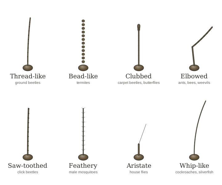
Antenna shape is one of the most useful clues for identification:
| Shape | Technical term | What it looks like | Examples |
|---|---|---|---|
| Thread-like | Filiform | Many similar segments, even width | Grasshoppers, ground beetles |
| Whip-like | Setaceous | Tapers steadily to a fine tip | Cockroaches, silverfish, mayflies |
| Bead-like | Moniliform | Round segments like a string of beads | Termites, some beetles |
| Saw-toothed | Serrate | Segments angled on one side | Click beetles and other beetles |
| Comb-like | Pectinate | Long branches on one side | Some beetles and sawflies |
| Clubbed | Clavate or capitate | Thickens toward the tip, gradually or suddenly | Butterflies, carpet beetles |
| Plate-like | Lamellate | Last segments form flat, fan-like plates | Some beetles |
| Elbowed | Geniculate | Sharp bend partway along | Ants, many weevils |
| Feathery | Plumose | Each segment carries long hairs | Some flies, including male mosquitoes |
| Aristate | Aristate | Short and pouch-like with one side bristle | House flies and relatives |
Compound eyes and ocelli
Most adult insects have a pair of compound eyes, each made of many small units called ommatidia. The count varies enormously: some worker ants have fewer than six, while some dragonflies have more than 25,000. Each unit samples one small part of the scene, and the brain builds an image from all of them.
Many insects also have two or three ocelli (simple eyes) on the top or front of the head. Ocelli do not form images. They respond to light level and quality and may help adjust the sensitivity of the compound eyes. Larvae of insects with complete metamorphosis, such as caterpillars and beetle grubs, lack compound eyes. Instead they have a few simple eyes called stemmata on each side of the head, which detect light and rough outlines.
Mouthparts
Chewing mouthparts are the starting design from which all other insect mouthparts evolved. A grasshopper shows the full set clearly.

From front to back, the parts are:
- Labrum: the upper lip, a flat plate hanging from the clypeus that holds food in place.
- Mandibles: a pair of hard jaws that move side to side to cut, crush or grind.
- Maxillae: a pair of second jaws behind the mandibles. Each has a base (cardo and stipes), two lobes (galea and lacinia) that work like a fork and spoon to handle food, and a jointed maxillary palp.
- Labium: the lower lip, formed from two appendages fused together. It closes the mouth from behind and carries a pair of labial palps.
- Hypopharynx: a tongue-like lobe in the middle of the mouth that helps mix food with saliva.
The palps on the maxillae and labium are sensory, used mostly for touch and taste.
Over time, many orders reshaped these same parts for liquid diets:
- Piercing-sucking (true bugs, aphids, mosquitoes): the parts are drawn out into fine stylets inside a sheath. In a female mosquito, six stylets (the mandibles, maxillae, labrum and hypopharynx) slide out of the labium to pierce skin.
- Siphoning (butterflies and moths): the two galeae of the maxillae join into a long tube that coils under the head and unrolls to drink.
- Sponging (house flies and blow flies): the labium ends in a spongy pad with fine grooves that draw up liquid.
- Chewing-lapping (honey bees): the mandibles stay as grasping jaws, while the maxillae and labium form a tongue for lapping nectar.
For a side-by-side view of each type, see insect mouthparts.
The thorax
The thorax is the locomotion center: it houses the big muscles that run the legs and wings. It has three segments, named from front to back:
- Prothorax: carries the first pair of legs. Its upper plate, the pronotum, is often enlarged into a shield, as in cockroaches, beetles and grasshoppers.
- Mesothorax: carries the second pair of legs and, in winged insects, the front wings.
- Metathorax: carries the third pair of legs and the hind wings.
Wings are never found on the prothorax. Each thoracic segment has an upper plate (notum), a lower plate (sternum) and side plates (pleura). In true bugs and beetles, part of the mesothorax shows from above as a triangular plate, the scutellum, between the wing bases.
Legs
Every insect leg has the same five main segments, joined by hinges:
- Coxa: the base, joined to the body.
- Trochanter: a small segment that links the coxa to the femur.
- Femur: usually the largest and strongest segment, holding most of the leg muscles.
- Tibia: a long, slender segment, often with spines.
- Tarsus: the “foot,” made of one to five small segments.
At the tip is the pretarsus, usually a pair of claws. Many insects add adhesive pads between or under the claws (the arolium or pulvilli), which help them grip smooth surfaces. The number of tarsal segments is a standard identification feature for beetles and many other groups.
Leg modifications
The same five segments are reshaped for very different jobs:
| Leg type | Technical term | How it is built | Examples |
|---|---|---|---|
| Running | Cursorial | Long, slender legs with spines | Cockroaches, ground beetles |
| Jumping | Saltatorial | Hind femur greatly enlarged and muscular | Grasshoppers, crickets, fleas |
| Digging | Fossorial | Front legs broad and shovel-like, with tooth-like points | Mole crickets |
| Grasping | Raptorial | Large, spiny front legs that seize and hold prey | Praying mantids |
| Swimming | Natatorial | Middle or hind legs flattened and fringed with stiff hairs, used as oars | Diving beetles, water boatmen |
| Clinging | (no single term) | Short tarsus ending in a single large claw for gripping hair | Sucking lice |
When running, insects use a tripod gait: the front and hind legs on one side move with the middle leg on the other side, so three legs always stay on the ground.
Wings
Most winged insects have two pairs, on the mesothorax and metathorax. Working wings appear only at the molt to adulthood. Mayflies are the one exception: they molt once more after gaining wings. A nymph’s wing pads or a larva’s lack of wings tells you it is not yet an adult.
Venation basics
A wing is a thin double layer of cuticle stiffened by tube-like veins. The veins carry blood (hemolymph), a breathing tube and a nerve. Entomologists name the main veins from the leading edge back: costa ©, subcosta (Sc), radius ®, media (M), cubitus (Cu) and the anal veins (A). Short crossveins connect them, and the enclosed spaces are called cells. Wing shape, texture and vein pattern are standard features for identifying insect groups, down to family and often genus.
In flight, the two pairs often work as one. Bees and wasps lock the hind wing to the front wing with a row of tiny hooks called hamuli.
Modified wings
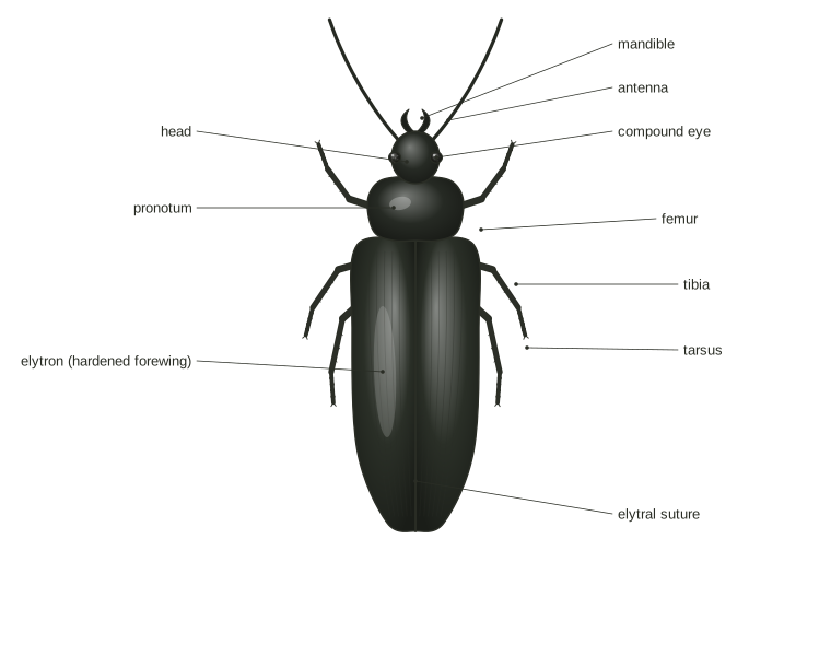
Front wings are often thickened to protect the hind wings, and the type of thickening identifies whole orders:
- Elytra (beetles): hard, shell-like front wings that meet in a straight line down the back and protect the membranous hind wings folded beneath them.
- Hemelytra (true bugs): front wings leathery near the base and membranous at the tip, crossing over the back in an X.
- Tegmina (grasshoppers, crickets, cockroaches, mantids, earwigs): front wings that are leathery all over, like parchment, covering fan-folded hind wings. Earwig tegmina are short.
- Halteres (true flies): the hind wings reduced to small knobbed stalks that vibrate in flight and act as balance organs.
- Scaled wings (butterflies and moths): both pairs covered in overlapping scales that hold the color patterns.
- Fringed wings (thrips): very narrow, rod-like wings edged with a dense fringe of long hairs.
- Hairy wings (caddisflies): covered in fine hairs rather than scales, one way to tell them from moths.
Some insects have lost their wings entirely. Silverfish never had them in their evolutionary history. Fleas, lice and worker ants come from winged ancestors and lost wings secondarily.
The abdomen
The abdomen holds most of the digestive tract, the heart, the reproductive organs and much of the breathing system. It has no walking legs in adult insects.
Segments
The ancestral insect had eleven abdominal segments. Silverfish and mayflies keep all or traces of them. In beetles and flies, segments are fused or tucked away so that as few as six or seven are visible. Each segment has a tergite above and a sternite below, joined by soft membranes.
Spiracles and tracheal breathing
Insects do not breathe through the mouth. Air enters through spiracles, small valved openings along the sides of the thorax and abdomen, usually one pair per segment. Many adults have a spiracle on each side of the first eight abdominal segments.
Each spiracle opens into the tracheae, a branching network of air tubes kept open by spiral bands of cuticle. The tubes divide into ever-finer branches that end at tracheoles next to individual cells, where oxygen dissolves and diffuses in. Small insects rely mostly on diffusion. Larger ones pump the abdomen to move air, opening some spiracles while closing others. Muscles can close the spiracles to save water in dry conditions.
Cerci
Many insects have a pair of cerci, sensory appendages at the tip of the abdomen. Earwig cerci are hardened into forceps. Silverfish and bristletails have two cerci plus a central tail filament, giving three “tails.” Mayflies have two or three tail filaments. Cerci are absent in true bugs, thrips, lice and all the orders with complete metamorphosis.
Ovipositor and stinger
Females of many insects have an ovipositor, an egg-laying tube built from appendages of abdominal segments 8 and 9. Grasshoppers, among others, use it to place egg pods in the soil.
In bees, ants and the stinging wasps, the ovipositor has become a stinger linked to venom glands. Because the stinger is a modified ovipositor, only females can sting; males cannot.
Males have external genitalia at the same end of the abdomen. Their shape often differs between closely related species, so specialists use them to confirm identifications.
Tympanal organs and other abdominal structures
Several insects hear with tympanal organs, thin, eardrum-like membranes. Grasshoppers and many moths have them on the abdomen. In grasshoppers they sit on the sides of the first abdominal segment (labeled tympanum in the hero illustration). Crickets and katydids have theirs on the front legs instead, just below the knee.
Other abdominal features worth knowing:
- Cornicles: a pair of short tubes on the back of most aphids that secrete fluids.
- Prolegs: fleshy, unjointed false legs on the abdomen of caterpillars and sawfly larvae. True legs are on the thorax only.
- Gills: leaf-like or thread-like plates on the abdomen of aquatic young such as mayfly and damselfly naiads.
Using anatomy to identify insects
Most first identifications rest on a handful of features you can see with a hand lens.
Count and inspect the wings. One pair plus halteres means a true fly. Hard covers meeting in a straight line mean a beetle. Half-leathery front wings crossing in an X mean a true bug. Scaled wings mean a butterfly or moth.
Check the antennae. Elbowed antennae point to ants and many weevils. Straight, bead-like antennae point to termites. Long, whip-like antennae are typical of cockroaches. A short antenna with a side bristle marks a house fly and its relatives.
Look at the waist. Ants, bees and wasps have a narrow, pinched waist. Termites and flies do not. This single feature settles the most common question about winged insects in homes in spring: see termite vs flying ant. Termite swarmers have straight antennae, a broad waist and two pairs of wings of equal length. Winged ants have elbowed antennae, a pinched waist and front wings longer than the hind wings.
Look at the body covering. Honey bees are hairier and less brightly striped than yellowjackets, which are boldly banded in black and yellow with a very narrow waist. See bee vs wasp for a fuller comparison.
Check the mouthparts. A beak folded under the body marks a true bug. A coiled tube marks a butterfly or moth. Visible jaws mean a chewing insect such as a beetle, cockroach or grasshopper.
Note the life stage. A wingless insect with wing pads is a nymph of an order with simple metamorphosis. A caterpillar has prolegs on the abdomen; a beetle grub does not.
Once you have the order, the orders of insects page lists its other features, and the identification hub narrows things down for insects found indoors.
References
- Exoskeleton (Bug Bytes, ENT 425). North Carolina State University.
- Head (Bug Bytes, ENT 425). North Carolina State University.
- Antennae (Bug Bytes, ENT 425). North Carolina State University.
- Mouthparts (Bug Bytes, ENT 425). North Carolina State University.
- Legs (Bug Bytes, ENT 425). North Carolina State University.
- Wings (Bug Bytes, ENT 425). North Carolina State University.
- Abdomen (Bug Bytes, ENT 425). North Carolina State University.
- Respiratory System (Bug Bytes, ENT 425). North Carolina State University.
- Acoustic Communication (Bug Bytes, ENT 425). North Carolina State University.
- Photoreceptors (Bug Bytes, ENT 425). North Carolina State University.
- Insect antennae. Amateur Entomologists' Society.
- Mole cricket. Texas A&M AgriLife Extension, Field Guide to Common Texas Insects.
- Subterranean Termites (ENTFACT-604). University of Kentucky Entomology.
See also
- The orders of insects
- Insect mouthparts
- Simple and incomplete metamorphosis
- Identify an insect
- Termite vs flying ant
This page was last updated on October 7, 2026. We research our guides from university extension services, government agencies, museums and peer-reviewed literature. Read the editorial policy or report an error.